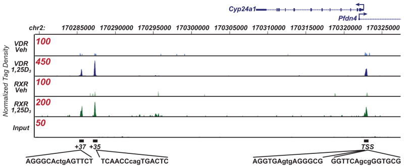
Figure 1.
ChIP-seq analysis of VDR and RXR binding at the mouse Cyp24a1 gene locus. MC3T3-E1 cells were treated with either vehicle or 1,25(OH)2D3 (10−7 M) for 3 hr and then subjected to ChIP-seq analysis using antibodies to either VDR or RXR. The genomic interval on mouse chromosome 2 and the location of the Cyp24a1 transcription unit (12 exons) including the direction of transcription (arrow) is shown at the top. An infrequently utilized promoter for the gene Pfdn4 and the direction of its transcription (not regulated by 1,25(OH)2D3) is also shown. Tracks indicate tag densities (normalized to 107 reads) for vehicle-or 1,25(OH)2D3-treated VDR or RXR binding. Note the scale for peak height is different for each track (shown in red) to highlight VDR and RXR peak activities. DNA sequences for the putative VDREs located promoter distal (two validated VDREs) and single VDREs at +35 and +37 kb found at the peak height are indicated below the tracks. TSS, transcriptional start site.

