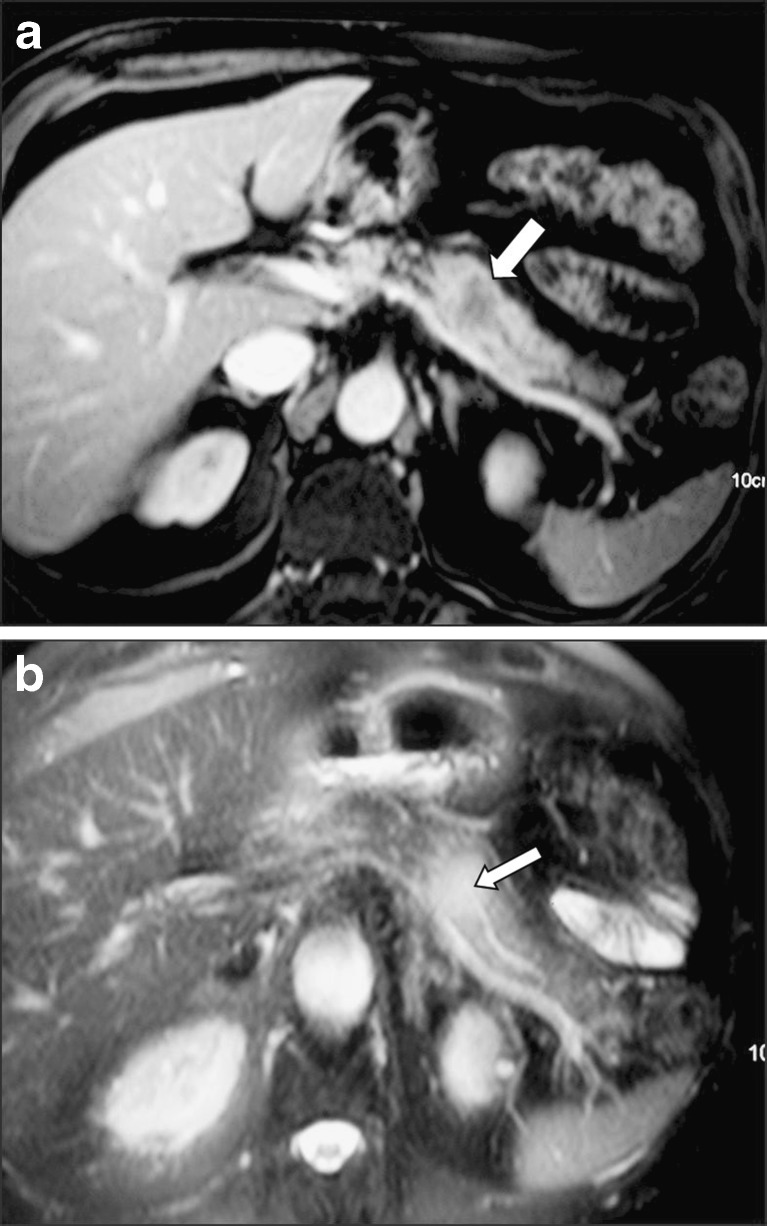
Fig. 10.
A 70-year-old man who underwent surgery for lung cancer. Six months later, he developed a solitary metastatic lesion in the pancreatic body evaluated by CT (arrow in a) and MRI (b). Note that the pancreatic duct “penetrates” the metastatic lesion with no evidence of infiltration on the T2-weighted image (arrow in b)

