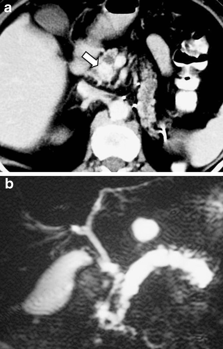
Fig. 9.
A 63-year-old woman who underwent left nephrectomy due to renal cell carcinoma. Three years later, the follow-up CT scan revealed a metastatic lesion with intense contrast enhancement in the uncinate process of the pancreas (arrow in a), causing dilatation of the main pancreatic duct in the body and tail that was evaluated by MRCP (b)

