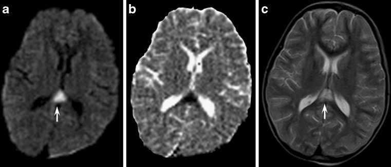
Fig. 10.
Axial MR images obtained in a child with a head injury at the level of the lateral ventricles. The midline of the splenium of the corpus callosum presented diffusion-weighted (a) and T2-weighted (c) hyperintensity (arrows) with reduced ADC values on ADC maps (b) consistent with diffuse axonal injury

