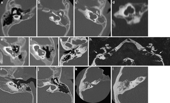
Fig. 3.

Inner ear dysplasias. a-d Patient 1: a 6-month-old girl presenting with congenital deafness has bilateral congenital malformations of the labyrinth on CT. On the right (a) axial CT shows a small cyst in place of the cochleovestibular system, corresponding to labyrinthine aplasia. The internal auditory canal (IAC) is also absent. On the left, axial (b, c) and coronal (d) CT shows an IAC bending posteriorly towards a cystic dilated vestibular system. There is no development of the cochlea anteriorly; hence this developmental anomaly can be classified as a cochlear aplasia. e, f Patient 2: on axial CT images of this deaf child, a widened cochlea without a modiolus renders a cystic appearance of the cochlea. The vestibule also shows pronounced enlargement. This has been classified as a cystic cochleovestibular dysplasia (incomplete partition type I). g, h Patient 3 shows a classic Mondini malformation (incomplete partition type II) consisting of a cochlea with a normal basal turn, but confluent middle and apical turn (black arrow), a widened vestibule (V) and an enlarged vestibular aqueduct (VA) (the normal size of the VA is 1–1.5 mm at its midpoint and generally does not exceed the size of the SCC). The modiolus is deficient (white arrowhead). On a 3D T2 TSE MR image the enlarged endolymphatic duct and sac are well seen (white arrows). i, j Patient 4: in this patient a foreshortened cochlea and a cystic appearance of the vestibular system is seen without enlargement of the vestibular aqueduct (black arrow). This dysplasia is more difficult to identify within the current classification system. k, l Patient 5: absence of the posterior semicircular canal illustrates that the abnormal development may be limited to the semicircular canals. Isolated absence of the posterior SCC may be seen in Waardenburg or Allagile syndrome
