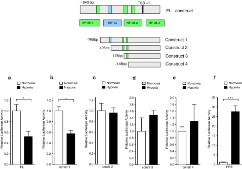
Fig. 4.
VASP promoter analysis during hypoxia in vitro. Schematic drawing of the putative VASP promoter. Displayed are the potential binding sequences for HIF-1α and NF-κB (green). Serial truncations were then performed relative to transcription start site (TSS) to identify the influence of HIF-1α or NF-κB on the putative VASP promoter HMEC-1 were transfected with the a VASP-PGL3 plasmid, b construct 1 excluding NF-κB binding site 1, c construct 2 excluding the HIF-1α binding site, d construct 3, e construct 4, and f HRE luciferase reporter driven by four tandem HREs (positive control) (Data are mean ± SEM, n = 5, * p < 0.05 as indicated).

