Fig. 5.
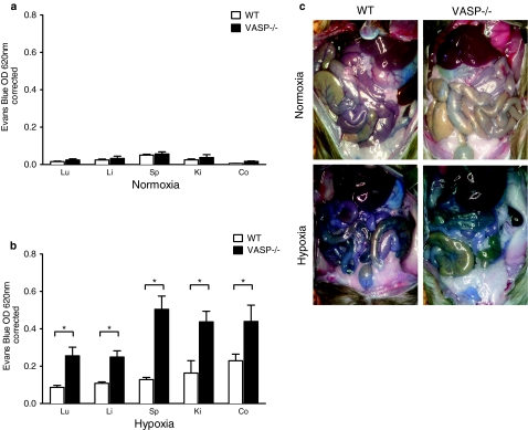
Increased vascular permeability in VASP −/− animals during hypoxia. WT and VASP −/− animals were injected with Evan blue dye and exposed to room air or normobaric hypoxia (8% O2, 92% N2) for 4 h. Animals were killed and the lung (Lu), liver (Li), spleen (Sp), kidney (Ki), and colon (Co) were harvested. Organ specific Evans blue concentrations were quantified and corrected for contamination by heme pigments at 740 nm. a Evans blue tissue extravasation in WT and VASP −/− animals following exposure for 4 h to normoxia. b Evans blue tissue extravasation in WT and VASP −/− animals following exposure for 4 h to hypoxia. c Representative images of abdominal dissections of WT and VASP−/− animals following exposure to normoxia or hypoxia for 4 h are demonstrated (All data are mean ± SEM, n = 8–9 per group, *p < 0.05 as indicated).

