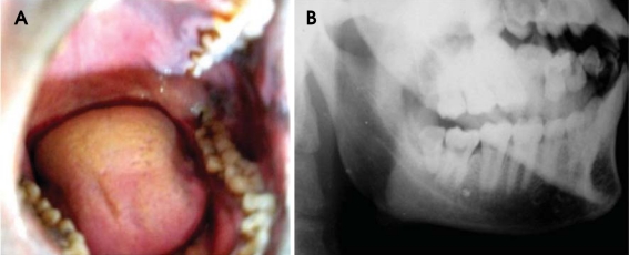
Fig. 1.
Case 1. A. Intraoral photograph shows a tooth-like structure with a blackish discoloration appeared to be embedded distal to the third molar. B. Lateral oblique radiograph reveals the presence of a partially impacted tooth having morphology similar to a molar distal to the third molar. The tooth is associated with a large radiolucency in the crown and a periapical lesion.

