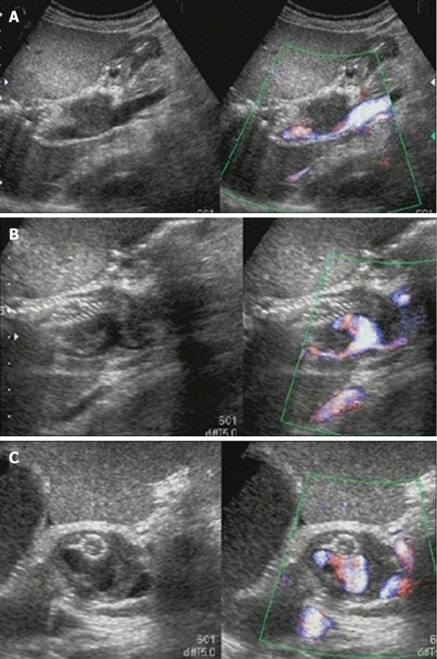
Figure 4.

Color Doppler ultrasonography on the 6th hospital day. A: Ultrasonography showed marked dilation of the common bile duct to a diameter of 24 mm and consequent extrinsic compression of the portal vein; B:The self-expandable metallic stent (SEMS) was displaced toward the liver, and hypoechoic solid components filling the space between the SEMS and bile duct and a pseudoaneurysm showing cystic growth in the lumen of the bile duct was observed; C: Only the apex of the pseudoaneurysm was in contact with the SEMS.
