
Figure 3.
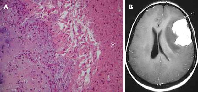
Gliosarcoma. A: Pathological image showing an area with features of osteogenic sarcoma (black asterisk) adjacent to the glial component of the tumor (white asterisk) (hematoxylin-eosin, original magnification × 100); B: On the axial, post-contrast T1-weighted magnetic resonance image, the tumor shows intense homogeneous enhancement. Note the close relation of the mass with the dura, which is enhanced in a way mimicking meningioma (arrow).