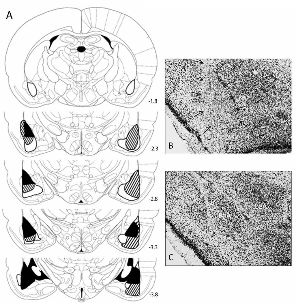
Figure 1.
(A) Drawings of smallest (dark shading), largest (light shading) and median lesions (lined) of basolateral amygdala (BLA), and photomicrographs of representative neurotoxic (B) and sham (C) lesions. In excitotoxic lesions (arrows), extensive neuron loss marked by gliosis is confined to the BLA, sparing the amygdala central nucleus (CeA). The numbers on the atlas sections refer to stereotaxic distances posterior to bregma. The sections are from The Rat Brain in Stereotaxic Coordinates (4th ed.), Figures 26, 29, 31, 33, 35, by G. Paxinos & C. Watson, 1998, New York: Academic Press. Adapted by permission of Elsevier.

