Figure 3.
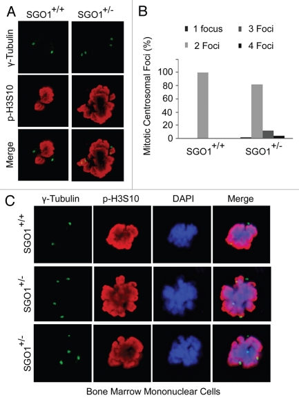
SGO+/− MEFs exhibit extra centrosomal foci. (A) Paired MEFs were stained with the antibodies to γ-tubulin (green) and p-H3S10 (red). DNA was stained with DAPI (blue). (B) Paired MEFs were stained with the antibody to γ-tubulin (green). Centrosomal numbers in mitotic cells were counted for each type of MEFs (n = 50 for each). (C) Bone marrow cells from mice of each genotype were stained with the antibodies to γ-tubulin (green) and p-H3S10 (red). DNA was stained with DAPI (blue). Representative images are shown.

