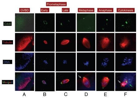
Figure 1.
Cdc6 is localized to spindle poles in MI. Stage VI oocytes were treated with progesterone to induce maturation, and samples were collected at various time and processed for immunofluorescence. Oocytes were stained for α tubulin (red), Cdc6 (green) and DNA (blue). Grey arrow in (D) indicates the animal cortex. Grey arrow in panel f indicates the first polar body and green arrow indicates the second meiotic spindle precursor. Image scale: 140 µm (A), 80 µm (B), 40–50 µm (CµF).

