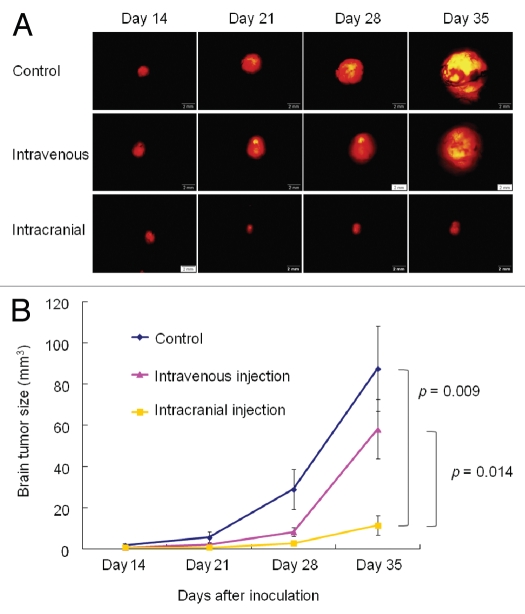
Figure 3.
Real-time, in vivo imaging of U87-RFP glioma growing orthotopically in nude mice and evaluation of S. typhimurium A1-R therapeutic efficacy over time. (A) Representative images of tumor growth in untreated group (control); treated with intravenous injection (i.v. injection); and treated by cranial injection of S. typhimurium A1-R (intracranial injection). Mice in the treatment groups were given S. typhimurium A1-R weekly by i.v. injection or intracranial injection for 3 weeks, beginning 14 d after tumor transplantation. Tumor size was measured using RFP imaging at days 14, 21, 28 and 35. Scale bars: 2 mm. (B) Tumor volumes in each group were compared. Tumor volume in the intracranial-injection group was significantly smaller than in the other two groups (at day 35, vs. control: p = 0.009; vs. intravenous: p = 0.014). However, there was no significant difference in tumor volume between the control group and intravenous-injection group. Seven mice were used in each group. The experimental data are expressed as the mean ± SD. Statistical analysis was performed using the Student t-test.

