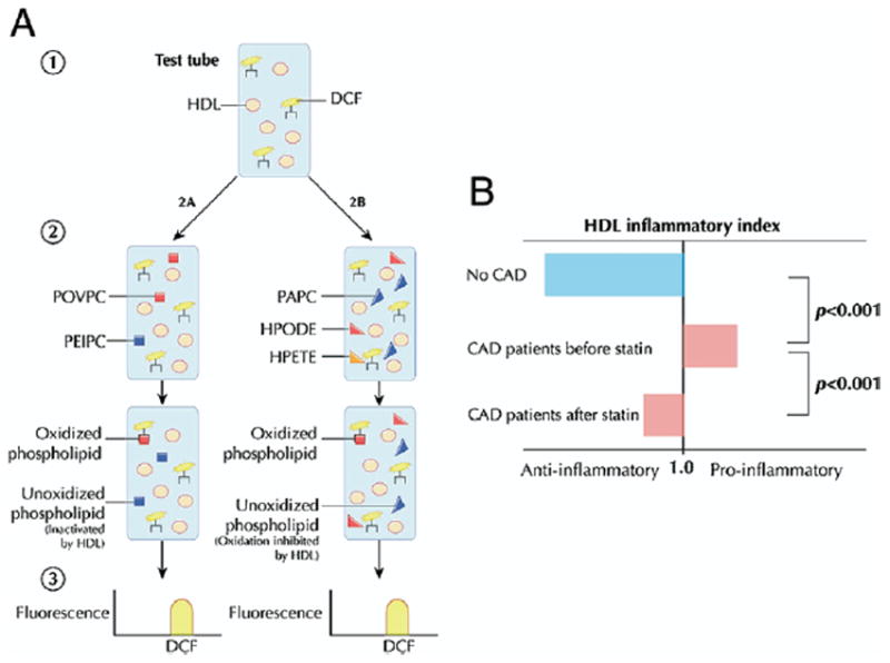
Figure 5. Lipid Oxidation.

(A) Dichlorofluorescein (DCF), a fluorescent marker of lipid oxidation products, is added to test HDL isolated from patient serum (85). Oxidized phospholipids found in mildly oxidized HDL, such as 1-palmitoyl-2(5-oxovaleroyl)-sn-glycero-3-phosphorylcholine (POVPC) and 1-palmitoyl-2(5,6-epoxyisoprostane E2)-sn-glycero-3-phosphorylcholine (PEIPC), are added to assess the ability of HDL to inactivate biologically active phospholipids. To assess the ability of HDL to prevent phospholipid oxidation, HDL is added to a mixture of 1-palmitoyl-2-arachidonoyl-sn-glycero-3-phosphorylcholine (PAPC) from unoxidized LDL and the endothelium-derived oxidants hydroperoxyoctadecadienoic acid (HPODE) or hydroperoxyeicosatetraenoic acid (HPETE). After the reagents are combined, spectroscopy permits quantification of net oxidation, with diminished fluorescence intensity signaling fewer oxidized phospholipids and suggesting a more antiatherogenic HDL. (B) The “inflammatory index” is derived by dividing net antioxidant activity in the presence of HDL by that observed in the absence of HDL. The HDL obtained from coronary artery disease (CAD) patients exhibits an impaired ability to antagonize monocyte chemotaxis and lipid oxidation compared with control subjects (75). The atheroprotective effect of HDL is partially restored following statin therapy. Figure illustrations by Rob Flewell. Abbreviations as in Figures 1 and 2.
