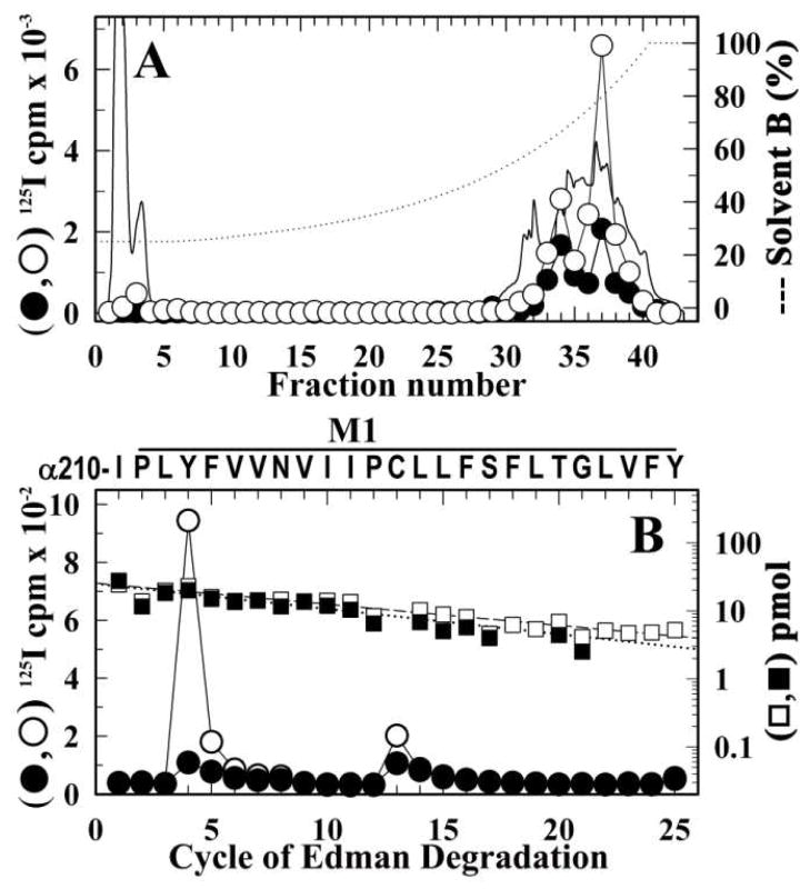
Figure 8. Identification of amino acids photolabeled by [125I]-SADU-3-72 in the α M1 segment of the resting Torpedo nAChR.
From the photoabeling experiment of Figure 6, α subunits from affinity-purified nAChR photolabeled in the presence of α-BgTx (●) or Carb (○) were digested ‘in-gel’ with V8 protease. The labeled fragment αV8-20K was then isolated and further digested with trypsin. The tryptic fragment αT5K was then isolated from a small-pore Tricine SDS-PAGE gel. Shown are reversed-phase HPLC fractionation (A) and amino acid sequence analysis (B) of the [125I]-SADU-3-72-labeled αT5K. A, The elution of peptides during HPLC was monitored by absorbance at 210 nm (solid line) and 125I elution was quantified by γ counting of each fraction (●, ○). B, 125I (●, ○) and PTH-amino acids (■, □) released during amino acid sequence analysis of 125I peak (fractions 36–38; ●, 3,000 cpm; ○, 10,000 cpm) from the HPLC purification of αT5K. In each sample, the primary peptide detected began at αIle210 (●, I0 = 25 ± 2 pmol, R = 92%; ○, I0 = 26 ± 2 pmol, R = 93%) and for both samples there were peaks of 125I cpm release in cycles 4 and 13 corresponding to labeling of αTyr213 (●, 1 cpm/pmol; ○, 10 cpm/pmol) and αCys222 (●, 2 cpm/pmol; ○, 3 cpm/pmol).

