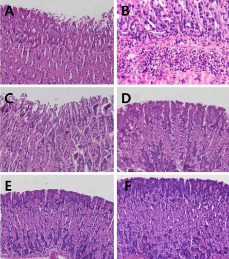
Figure 1.
Representative microscopic findings of the gastric mucosa of mice infected with H. pylori followed by treatment with IgY (50-200 mg/kg) or pantoprazole (30 mg/kg). Note the degeneration and sloughing of villi (A) and submocosal inflammatory cell infiltration (B) in vehicle group, in comparison with light sloughing of villi at a low (50 mg/kg) dose of IgY (C) and near-normal features at high doses of IgY (D, 100 mg/kg; E, 200 mg/kg) or pantoprazole (F, 30 mg/kg).

