Figure 9.
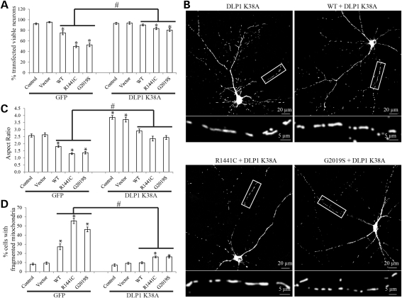
Dominant-negative DLP1 rescued LRRK2-induced mitochondrial abnormalities in primary neurons. Rat E18 primary cortical neurons (DIV = 7) were transiently co-transfected with myc-tagged LRRK2 (WT, R1441C or G2019S), GFP/GFP-tagged DLP1 K38A and mito-DsRed2 at a ratio of 9:1:1. (A) Quantification of neuronal viability in positive transfected neurons 3 days after transfection. Mitochondrial morphology was evaluated 2 days after transfection (B–D). (B) Representative pictures show that co-overexpression of DLP1 K38A mutant prevents LRRK2-induced mitochondria fragmentation in rat E18 primary cortical neurons 2 days after transfection. The boxed area was enlarged immediately below the picture. (C and D) Quantification of mitochondria morphology in primary neurons transfected with indicated plasmids. At least 20 cells were analyzed in each experiment and experiments were repeated three times (asterisk represents P< 0.05 when compared with the control neurons and hash symbol represents P< 0.05 when compared with neurons with only LRRK2 overexpression; Student's t-test).

