Figure 2.
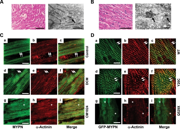
Histopathology and comparative immunohistology of an explanted human heart from siblings with RCM and NRCs. (A) Histology (left panel, bar = 100 μm) and electron micrograph (right panel, bar: 3 μm) of the LV myocardium from patient CM1023 with RCM. An arrow indicates electron-dense material accumulated in the intercellular junctions. (B) Histology (left panel, bar = 100 μm) and electron micrograph (right panel, bar: 3 μm) of the LV myocardium from patient CM1024 with RCM. Arrows indicate electron-dense material accumulated in the intercellular junctions similar to that seen in the sibling CM1023. (C) Expression of MYPN (a, d and g), α-actinin (b, e and h) and merged images (c, f and i) in human control heart (a–c), DCM (d–f) and RCM (CM1024, g–i) patients. Bar = 10 μm. Arrows indicate co-localized MYPN and α-actinin at the striated Z-discs. Asterisks indicate disrupted sarcomeric Z-discs with abnormal diffuse MYPN co-distributed focally with α-actinin in CM1024. (D) Expression of α-actinin (red) in NRCs transfected with GFP-WT-MYPN (a–c), GFP-Y20C-MYPN (d–f) or GFP-Q529X-MYPN (g–i). Right panels represent merged images. Arrows indicate GFP-WT-MYPN and GFP-Y20C-MYPN co-localized with α-actinin. Asterisks indicate disorganized GFP-Q529X-MYPN in the Z-discs and its disrupted co-localization with α-actinin. Bar = 5 μm.

