Figure 4.
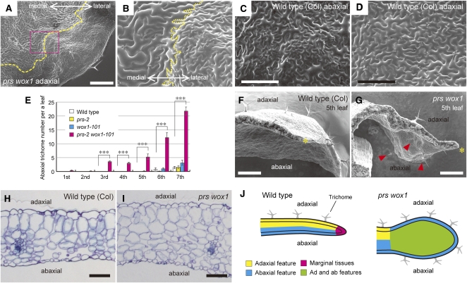
Adaxial/Abaxial Patterning of Tissue Differentiation in the Wild-Type and prs wox1 Leaves.
(A) and (B) Scanning electron micrographs of the adaxial surface of prs wox1. Yellow dashed lines, the boundary between the adaxial-type epidermis and abaxial-type epidermis.
(C) and (D) Scanning electron micrographs of the abaxial (C) and adaxial (D) surfaces of the wild-type leaves.
(E) The number of abaxial trichomes. Data are represented as the means ± sd. ***P < 0.001 by Tukey’s HSD test. In total, 11 wild-type and prs-2 and 9 wox1-101 and prs-2 wox1-101 samples were assessed.
(F) and (G) Scanning electron micrographs of the wild-type (F) and prs wox1 leaves (G) in cross section. Asterisks, leaf edges; arrowheads, abaxial trichomes.
(H) and (I) The medial region of the eighth leaves in cross section in the wild type (H) and prs wox1 (I).
(J) Schematic view of leaf margin phenotypes.
Bars = 500 μm in (A), (F), and (G), 200 μm in (C) and (D), and 100 μm in (H) and (I).

