Figure 2.
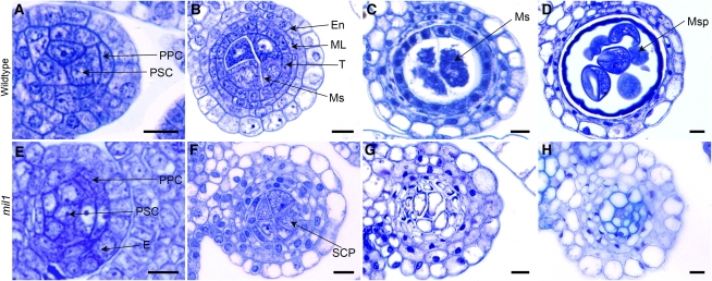
Toluidine Blue–Stained Transverse Sections of Anthers in the Wild Type and mil1.
(A) to (D) The wild type.
(E) to (H) mil1.
(A) and (E) Anthers with primary parietal cells and primary sporogenous cells.
(B) Wild-type anthers with microsporocytes and four parietal layers.
(C) Wild-type anthers at meiotic stage exhibiting crushed middle layer.
(D) Wild-type anthers filled with microspores, with middle layer and tapetum degraded.
(F) mil1 anthers with SCPs in the center. They generally have four parietal layers but show extra layers in some regions.
(G) mil1 SCPs increase in number and decrease in size as anthers develop. No cell degeneration is observed in wall cells.
(H) mil1 anthers are composed of somatic cells.
En, endothecium; ML, middle layer; Ms, microsporocyte; Msp, microspore; PPC, primary parietal cell; PSC, primary sporogenous cell; T, tapetum. Bars = 10 μm.
[See online article for color version of this figure.]

