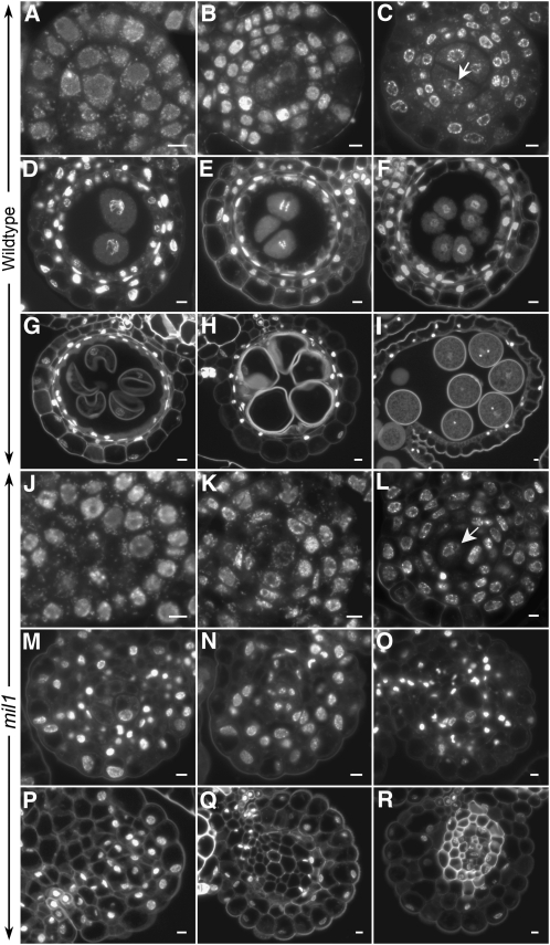
Figure 3.
DAPI-Stained Transverse Sections of Anthers in Both the Wild Type and mil1.
(A) to (I) The wild type.
(J) to (R) mil1.
(A) and (J) Anthers with sporogenous cells and primary parietal cells.
(B) and (K) Anthers with two secondary parietal cells.
(C) and (L) Anthers with four parietal cells established. Wild-type microsporocytes (arrow in [C]) are in premeiotic interphase, while mil1 SCPs (arrow in [L]) behave like somatic cells.
(D) and (E) Wild-type anthers with microsporocytes in meiosis. The middle layer is crushed.
(F) Wild-type anthers with tetrads formed.
(G) to (I) Wild-type anthers showing developing microspores. The tapetum is degenerated.
(M) to (R) mil1 SCPs show no sign of meiosis, and together with anther wall cells they develop into somatic cells.
Bars = 5 μm.

