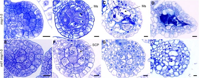
Figure 6.
Toluidine Blue–Stained Transverse Sections of Anthers in Both msp1-5 and the mil1 msp1-5 Double Mutant.
(A) to (D) msp1-5.
(E) to (H) mil1 msp1-5.
(A) and (E) msp1-5 and mil1 msp1-5 anthers develop normally at early developmental stage, with primary parietal cells and primary sporogenous cells.
(B) and (F) msp1-5 and mil1 msp1-5 both have excess microsporocytes (or SCPs) and almost two parietal layers.
(C) and (D) msp1-5 anthers have degraded microsporocytes, but intact parietal cells remained.
(G) and (H) mil1 msp1-5 SCPs behave like mil1 SCPs, increasing in number and decreasing in size as anthers develop, finally developing into somatic cells.
Ms, microsporocyte. Bars = 10 μm.
[See online article for color version of this figure.]

