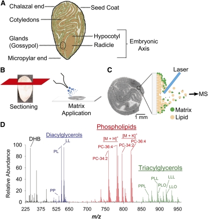
Figure 1.
Schematic Overview of Cottonseed Anatomy and the MALDI-MSI Process.
(A) Longitudinal diagram of a mature cotton embryo primarily composed of folded cotyledon tissues surrounding the embryonic axis (hypocotyl and radicle). Pigment glands containing the terpenoid gossypol are present throughout the tissue.
(B) Intact embryos are chemically fixed and sliced into thin cross or longitudinal sections with a cryostat. Sections of ~30 μm are adhered to a glass slide and coated with matrix, 2,5-dihydroxybenzoic acid (DHB).
(C) A laser beam is rastered across the section in a serpentine pattern ionizing the seed lipid/matrix mixture at an imaging resolution of 50 μm. Generated lipid ions as [M + H]+, [M + Na]+, or [M + K]+ adducts are directed into a mass analyzer for the mass spectral data acquisition.
(D) Each laser shot and detection cycle generates a raw data spectrum for each x, y position of the seed section. A sample spectrum shows the identification of the matrix (DHB identified as [2DHB − 2H2O + H]+ at m/z 273.04) and three distinct compound regions, phospholipids, and in particular PC (red region), TAGs (green region), and diacylglycerols (blue region) from in-source fragmentation of TAG and PC species. Representative species and adducts are labeled with their total acyl composition. For a more comprehensive list of species and adducts identified, see Supplemental Tables 1 to 4 online.

