Figure 3.
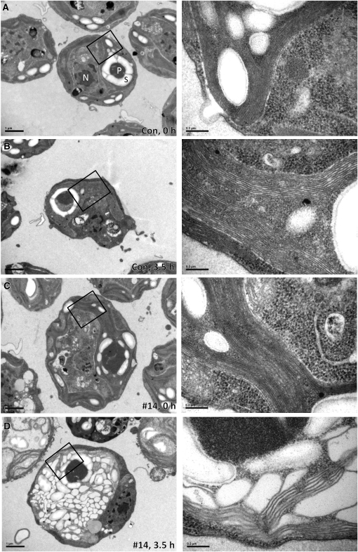
Thylakoids in VIPP1-amiRNA Strains Exposed to High Light Intensities Are Extremely Swollen.
(A) Electron microscopy image of a cell from the control strain grown at low light intensities. Cells were grown at ~30 μE m−2 s−1 in TAP-NH4 medium. An overview image is shown on the left, and a zoom-in of the region demarcated by the black box is shown on the right. N, nucleus; P, pyrenoid; S, starch. Bars in overview images correspond to 1 μm and those in zoom-ins to 0.2 μm.
(B) Electron microscopy image of a cell from the control strain exposed to high light. Cells were grown at ~30 μE m−2 s−1 in TAP-NH4 medium and exposed to ~1000 μE m−2 s−1 for 3.5 h. Images were taken as in (A).
(C) Electron microscopy image of a cell from a VIPP1-amiRNA strain grown at low light intensities. VIPP1-amiRNA strain #14 was grown at ~30 μE m−2 s−1 in TAP-NH4 medium. Images were taken as in (A).
(D) Electron microscopy image of a cell from a VIPP1-amiRNA strain exposed to high light. VIPP1-amiRNA strain #14 was grown at ~30 μE m−2 s−1 in TAP-NH4 medium and exposed to ~1000 μE m−2 s−1 for 3.5 h. Images were taken as in (A).

