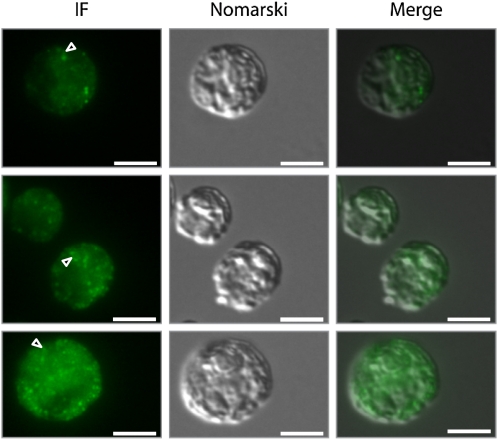
Figure 9.
Immunofluorescence Microscopy Detects VIPP1 in Distinct Punctae and as Diffuse Material in the Chloroplast.
Control cells from the cw15-325 background were grown at ~30 μE m−2 s−1 in TAP-NH4 medium and fixed and processed for immunofluorescence (IF) microscopy as described in Methods. The signal recognized by the affinity-purified anti-VIPP1 antibody is shown in green. Triangles indicate potential rod-like extensions. Bars = 5 μm.

