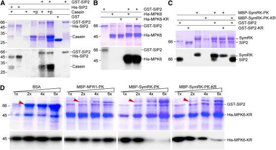
Figure 3.
In Vitro Kinase Activity of SIP2.
(A) Protein kinase activity of SIP2. SIP2 was expressed as a GST fusion protein in vector pGEX-6P1. GST alone served as a control. SIP2 was also expressed as a His tag fusion protein in pET28a. Affinity-purified GST, GST-SIP2, and His-SIP2 were incubated with casein or calf intestinal phosphatase–treated casein in the presence of [γ-32P]ATP. The reaction products were analyzed in SDS-PAGE. The gel was stained with Coomassie blue (top), dried, and subjected to autoradiography (bottom). Molecular mass standard (kD) is shown on left side.
(B) Phosphorylation of MPK6 MAPK by SIP2. Purified GST-SIP2 was incubated with Arabidopsis MPK6 or its kinase-negative mutant, MPK6-KR, in the presence of [γ-32P]ATP. Kinase reaction products were resolved on SDS-PAGE. The gel was stained with Coomassie blue (top) and subjected to autoradiography (bottom).
(C) SIP2 and SymRK did not phosphorylate each other. Because both SIP2 and SymRK could undergo autophosphorylation, their kinase-negative mutants were used as substrates. SIP2 and its kinase-negative mutant were purified on GST beads, whereas SymRK and its kinase-negative mutant were isolated via the MBP tag using chitin beads. In the presence of [γ-32P]ATP, the kinase-negative SymRK-PK-KR was not phosphorylated by SIP2. Similarly, the kinase-negative SIP2-KR was not phosphorylated by SymRK-PK.
(D) SymRK inhibited the phosphorylation of MPK6 by SIP2. The kinase-negative MPK6-KR was purified on Ni beads and used as substrate for SIP2 in the presence of [γ-32P]ATP. An increasing amount of SymRK-PK or its kinase-negative SymRK-PK-KR was added to the reaction mix. Increasing amounts of BSA and purified NFR1-PK were used to test the specificity of inhibition. The base amount (1×) of effector proteins (BSA, NFR1, and SymRK) was 1.0 μg per reaction, and the increasing amounts are indicated as relative quantities (2× to 6×). The bands corresponding to the increased amount of added proteins are labeled by arrows. The reaction products were analyzed on SDS-PAGE gels stained with Coomassie blue (top). Autoradiographs show the corresponding phosphorylated MPK6-KR (bottom).
[See online article for color version of this figure.]

