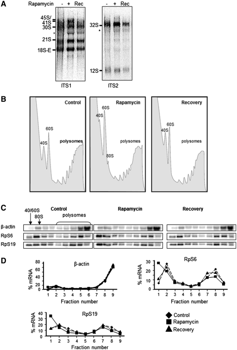
Figure 4.
Effect of rapamycin and its removal on ribosome distribution. HeLa cells were incubated with 100 nM rapamycin for 4 h and then the medium was replaced with fresh medium without the inhibitor for 2 h before the lysis. (A) Total RNA was extracted and subjected to northern blot analysis for pre-rRNA species. The membrane was hybridized with probes ITS1, ITS2, 18S and 28S as previously described. (B) Polysomal profile after rapamycin removal of HeLa cells. The polysomal and non-polysomal fractions are indicated. (C) Northern blot analysis of cytoplasmic RNA extracts from each fraction of sucrose gradient and analyzed with indicated probes (RpS6, RpS19 and β-actin). (D) Quantification of the signals is reported as a linear plot of percentage of mRNAs in each fraction.

