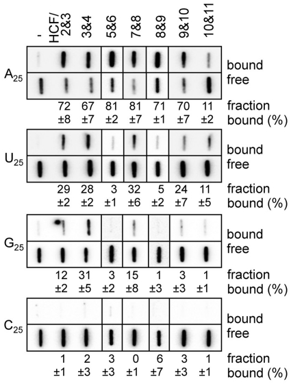
Figure 6.
Nucleobase preference of the mini-PPR proteins. The filter binding assay was conducted with ribonucleotide homo-polymer (A25, U25, G25 and C25; 250 pM) and the indicated mini-PPR protein (200 nM). Samples were filtered through the nitrocellulose and nylon membranes layer. The protein–RNA complexes were captured on the nitrocellulose membrane (bound). RNA that passed through the nitrocellulose was retained on the underlay of nylon membrane (free). Averages of the ratio of protein–RNA complexes (fraction bound, %) and the standard deviation (n ≥ 3) are shown.

