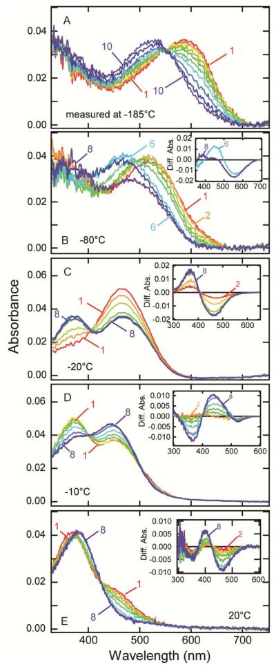
Figure 5.
Photobleaching process of lizard parietopsin. (A) Thermal reactions of bathoparietopsin. The photo-steady-state mixture mainly containing batho intermediate (curve 1) was warmed to −170 °C and recooled to −185 °C for the measurement of the spectrum (curve 2). Similarly the sample was warmed to −160, −150, −140, −130, −120, −110, −100 and −90 °C in a stepwise manner, and the spectrum was recorded at −185 °C after each warming (curves 3-10). (B) After the measurements shown in panel (A), the absorption spectrum of the sample was recorded at −80 °C (curve 1). It was warmed to −70, −60, −50, −40, −30, −20 and −10 °C in a stepwise manner, and the spectra were measured at −80 °C (curves 2-8). Inset: the difference spectra calculated by subtracting curve 1 from curves 6 and 8. (C) Transition from metaparietopsin-I to metaparietopsin-II at −20 °C. Parietopsin sample was irradiated with >560 nm light (20 mW/cm2) for 15 sec. The spectra were recorded at 1.5, 4.5, 9, 16, 32, 60, 120 and 150 min after irradiation (curves 1-8). Inset: the difference spectra calculated by subtracting curve 1. (D) Transition from metaparietopsin-II to metaparietopsin-III was observed at −10 °C. Parietopsin sample was irradiated with >560 nm light (20 mW/cm2) for 15 sec. The spectra were recorded at 1.5, 4.5, 9, 16, 32, 60, 120 and 150 min after irradiation (curves 1-8). Inset: the difference spectra calculated by subtracting curve 1. (E) Dissociation of metaparietopsin-II and metaparietopsin-III into retinal and opsin at 20 °C. Parietopsin sample was irradiated with >500 nm light (27 mW/cm2) for 20 sec. The spectra were recorded at 1.5, 5, 10, 15, 25, 60, 120 and 200 min after irradiation (curves 1-8). Inset: the difference spectra calculated by subtracting curve 1.

