Figure 6.
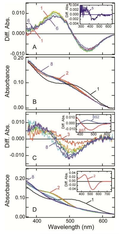
Photoreaction of detergent free parietopsin in membranes. (A) Transient absorption spectra of parietopsin in HEK293T membrane fragments at −20 °C. The sample was prepared by the sucrose flotation method and added 50% (w/v) glycerol. The spectra were recorded at 1.5, 5, 10, 30, 60 and 120 min after irradiation with >560 nm-light (20 mW/cm2) for 15 sec. Difference spectra was calculated by subtracting the spectrum recorded before irradiation from the spectra after irradiation (curves 1-6). Inset: the difference spectra between curve 1 (1.5 min) and curve 5 (60 min). (B) Transition from metaparietopsin-I to metaparietopsin-II plus metaparietopsin-III in PC liposomes. Parietopsin in PC liposomes (curve 1) was irradiated with a 532-nm laser pulse at 0 °C. The spectra recorded at 0.01, 0.05, 0.5, 1, 60, 600, 1800 sec after irradiation (curve 2-8) are displayed. (C) Difference spectra calculated by subtracting curve 2 from curves 3-8 shown in (B). Inset: Two b-spectra (BS1 and BS2) calculated from the spectral change shown in Figure 6C. BS1 and BS2 indicate transitions from metaparietopsin-I to metaparietopsin-II and from metaparietopsin-II to metaparietopsin-III, respectively. (D) Transition from metaparietopsin-II and metaparietopsin-III to retinal and apoprotein in PC liposomes. Parietopsin in PC liposomes (curve 1) was irradiated with >560 nm light (20 mW/cm2) for 5 min at 0 °C. The spectra were recorded at 5, 30 and 60 min and 2, 5, 10 and 14 hours after irradiation (curves 2-8). Inset: the difference spectra between curve 1 and curve 2, showing that metaparietopsin-II and metaparietopsin-III are formed by irradiation.

