Figure 5.

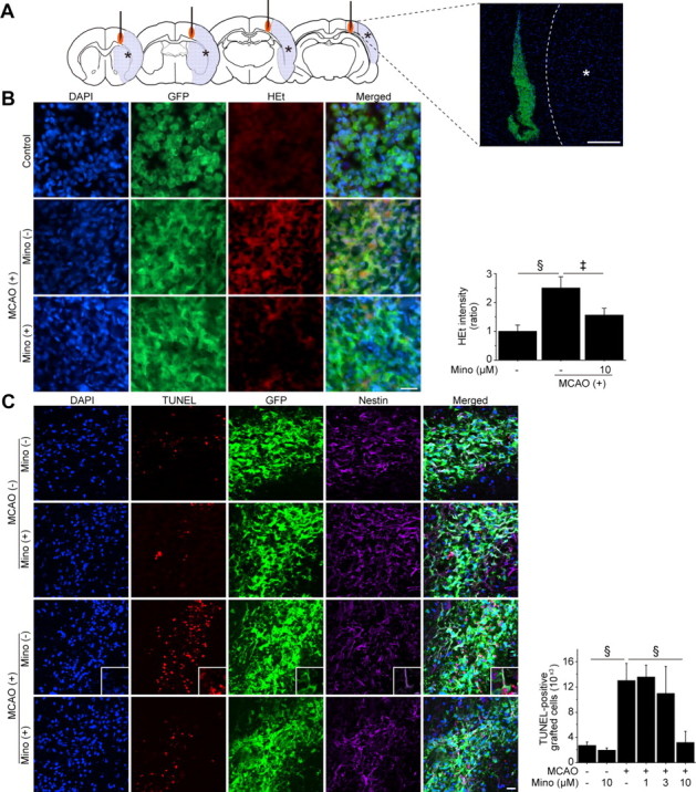
Reduced grafted-cell death with minocycline preconditioning in vivo. A, A schematic diagram and fluorescent staining with DAPI (blue) and GFP (green) 1 h after transplantation show the location of the graft and the ischemic lesion (*). The NSCs, with or without minocycline preconditioning, were transplanted 6 h after stroke. Scale bar, 200 μm. B, Fluorescent staining with DAPI (blue), GFP (green), and HEt (red) in brain sections 1 h after transplantation. HEt signals increased in the non-PCNSCs under ischemic reperfusion injury, but this signal increase was significantly reduced in the PCNSCs (10 μm) (n = 4). Scale bar, 20 μm. MCAO, Middle cerebral artery occlusion; Mino, minocycline. C, Fluorescent staining with DAPI (blue), TUNEL (red), GFP (green), and nestin (magenta) 2 d after transplantation. The number of TUNEL-positive grafted cells was similar in the non-PCNSC and PCNSC groups in the intact brain. When the non-PCNSCs were transplanted into the ischemic brain, the number of TUNEL-positive grafted cells increased remarkably. Minocycline preconditioning (10 μm) significantly reduced the number of TUNEL-positive grafted cells. The insets represent high magnification images showing the colocalization of TUNEL with nestin- and GFP-positive grafted cells (n = 4). Scale bar, 20 μm. ‡p < 0.005; §p < 0.001.
