
Fig. 1.
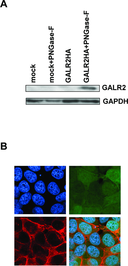
Exogenous GALR2 expression in transfected UM-SCC-1 cells. A, Immunoblotting shows exogenous GALR2 expression in pCMVGALR2IresGFP-transfected cells detected using antibody to HA-tag. Lanes labeled mock and GALR2HA contain protein lysates from mock-transfected and GALR2HA-transfected UM-SCC-1 cells (UM-SCC-1-GALR2). Lanes labeled mock+F and GALR2HA+F contain protein from cell lysates that were digested with N-glycosidase F. B, Exogenous GALR2 localizes to the cell membrane in UM-SCC-1-GALR2 cells. UM-SCC-1-GALR2 cells were stained with mouse monoclonal anti-HA-tag antibody and Hoechst 33342. Photographs show cells stained with Hoechst 33342 (upper left), GFP (upper right), HA-tag (lower left), and the merged image (lower right) (magnification × 400).