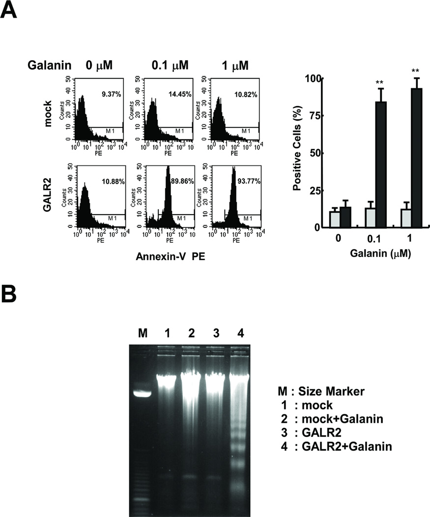
Fig. 5.

Induction of apoptosis in galanin-treated UM-SCC-1-GALR2 cells. A, Annexin-V staining. The annexin-V-positive cells were stained and counted by flow cytometry (left). Percent positive is the ratio of annexin-V-positive cells to total cell number (right) (**P<0.01). B, DNA fragmentation analysis. Genomic DNA was isolated and electrophoresed on a 2 % agarose gel. The DNA ladders were visualized under UV light with ethidium bromide. Lane M: Size marker. Lane 1: DNA isolated from UM-SCC-1-mock cells without galanin treatment. Lane 2: UM-SCC-1-mock cells with galanin treatment. Lane 3: UM-SCC-1-GALR2 cells without galanin treatment. Lane 4: UM-SCC-1-GALR2 cells with galanin treatment.
