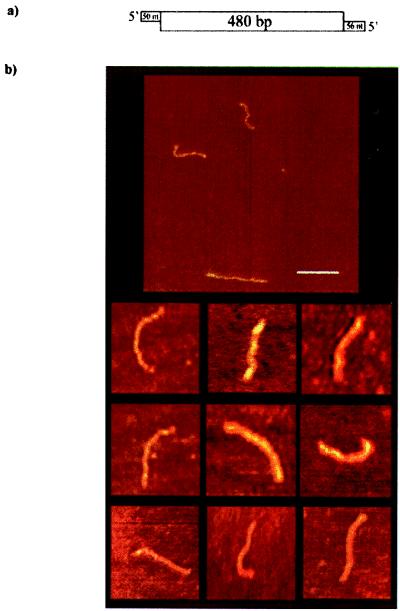
Figure 2.
AFM images of free dsRNA. (a) Schematic of the RNA construct used for the AFM imaging containing two 5′ (≈50-nt) single-stranded overhangs at both ends of the molecule. The double-stranded central region (480 bp) was generated by hybridization of the complementary regions of both strands. (b) AFM images of long dsRNA in air on mica. (Bar = 100 nm.) The rod-shaped molecules possess a mean end-to-end contour length of 134.5 ± 4 nm, which is consistent with an A form dsRNA.

