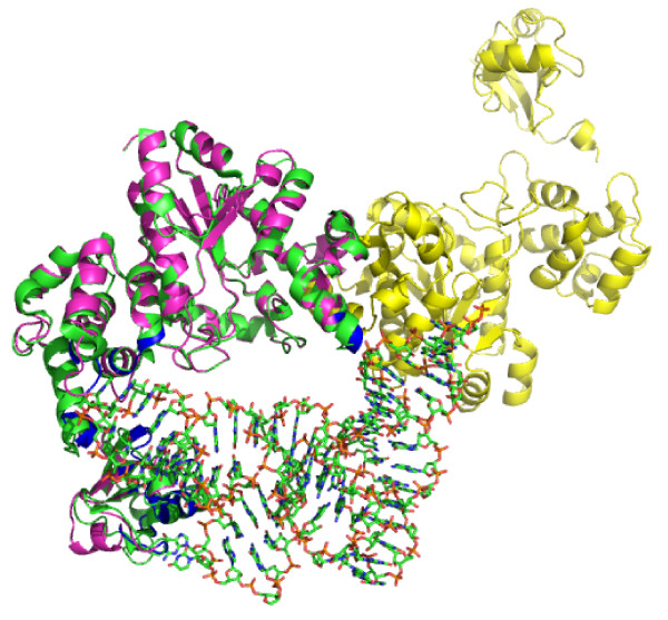
Figure 6.
TyrS structural model achieved using Swiss-Pdb Viewer v. 4.04 software and structure superposition onto the highly similar Thermus terhmophilus tyrosyl-tRNA synthetase. (Protein Data Bank: 1H3E). 1H3E is shown in green, and TyrS model is shown in magenta and yellow. Analysis of the two aligned structures indicates that all of the DNA/RNA binding sites are in regions that interact with tRNA in the 1H3E structure (shown in blue).

