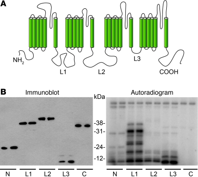
Figure 3. PKCε phosphorylates the third intracellular loop of Nav1.8.
(A) Schematic diagram illustrating the structural topology common to all eukaryotic sodium channels. (B) Intracellular domains of NaV1.8 were expressed in bacteria as 6xHis-tagged fusion proteins, and their expression was confirmed by Western blot analysis (left) with an anti-6xHis antibody (N terminus [N], ~24 kDa; L1, ~38 kDa; L2, ~39 kDa; L3, ~11 kDa; C terminus [C], ~35 kDa). Fusion proteins were used in a PKCε assay to determine whether any were PKCε substrates (right). An autoradiogram illustrates that the L3 loop (~11 kDa) is a likely PKCε substrate. Similar amounts of each fusion protein were used in Western blots (left) and kinase assays (right).

