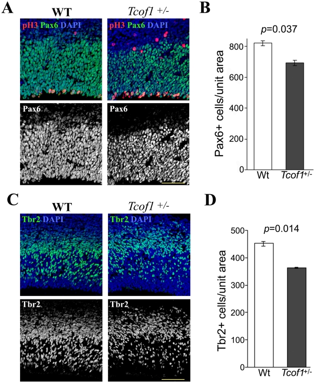
Figure 3. Tcof1 deficiency affects the number of Pax6-positive apical progenitors and Tbr2-positive basal progenitor cells in the telencephalon.
(A) Immunofluorescence detection of Pax6-positive progenitor cells (green) and anti-phospho-Histone H3 antibody (red) mitotic cells in the telencephalon of E14.5 wild-type and Tcof1 +/− embryos. Tissue sections were counterstained with DAPI (blue). (B) Bar graph depicting the number of Pax6-positive cell in the wild-type and Tcof1 +/− brain in a unit section of 125 µm width. (C) Co-immunostaining of the neuroepithelium of E14.5 wild-type and Tcof1 +/− embryos for Tbr2-positive neurons (green) and pH 3-positive (red) mitotic cells (D). Bar graph quantifying the number of Tbr2-positive cells in Tcof1 +/− embryos and their wild-type littermates in a unit section of 100 µm width. Scale Bars: A and C, 20 µm.

