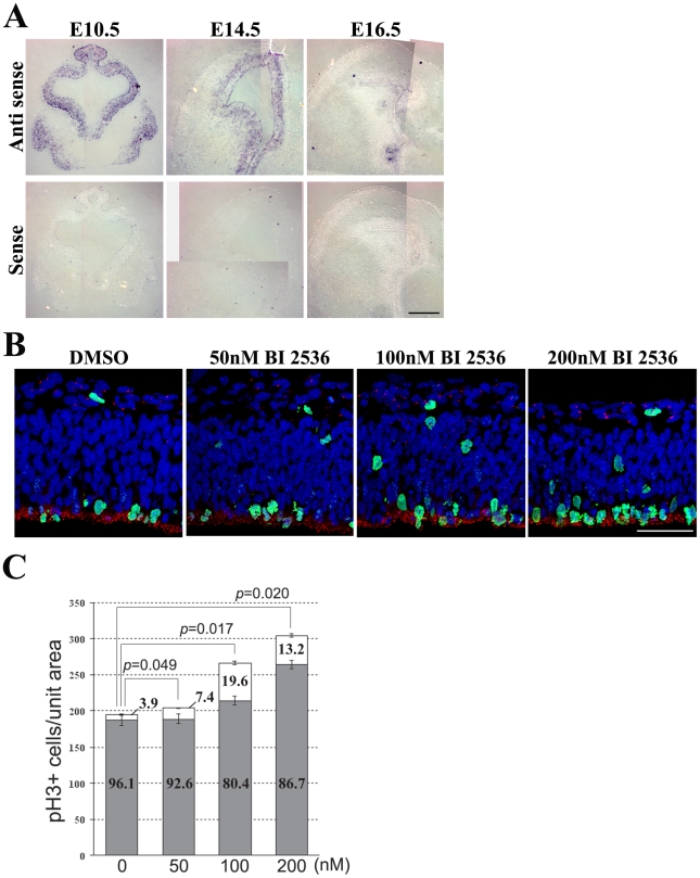
Figure 8. Plk1 co-operates in controlling mitotic progression and mitotic spindle orientation.
(A) Expression of Plk1 mRNA detected by in situ hybridization on coronal sections of E10.5–E16.5 embryos. (B) E11.0 mouse embryos were cultured with 50–200 nM BI 2536 for 16 hours. Mitotic cells were analyzed by immunostaining with phospho-Histone H3 (green) and Centrin (red). (C) The number of total pH 3-positive cells and percentage of surface and non-surface mitotic cells were quantified. The nuclei were stained with DAPI (blue). Scale Bars: A, 200 µm; B, 50 µm.

