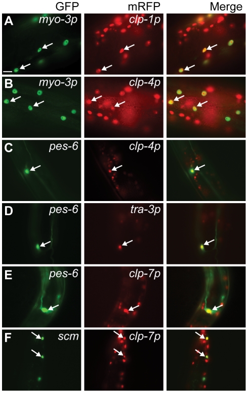
Figure 3. Calpain nls::mrfp expression in non-neuronal tissues.
Co-localization of the myo-3p::gfp::nls body wall muscle reporter with (A) clp-1p::nls::mrfp (crEx65) and (B) clp-4p::mrfp (crEx74). Co-localization of the pes-6::gfp excretory cell reporter with (C) clp-4p::nls::mrfp (crEx74), (D) tra-3p::nls::mrfp (crEx78) and (E) clp-7p::nls::mrfp (crEx79). (F) Co-localization of the scm::gfp seam cell reporter with clp-7p::nls::mrfp (crEx79). Tissue-specific GFP reporter, left (green), calpain promoter driven nls::mrfp expression, middle (red) and co-localization, right (yellow) highlighted with arrows. Each micrograph is typical of the pattern observed with at least two other independent transgenic strains generated with the same reporter construct. Scale bar, 10 µM.

