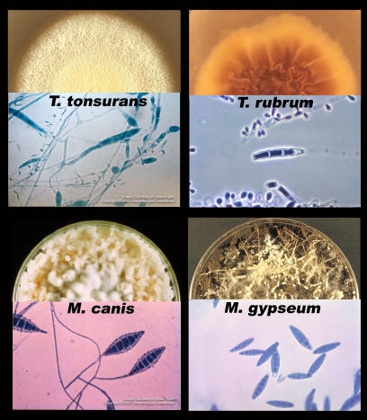
Figure 2. Major dermatophyte species—appearance in the laboratory.
Each section shows one of four species of dermatophyte, including a semicircle of fungus growing on an agar plate (top) and a microscopic picture of the asexual spores (macroconidia and microconidia, bottom). Photos courtesy of Doctor Fungus (http://www.doctorfungus.org) and the Public Health Image Library (PHIL, http://phil.cdc.gov/phil/home.asp).

