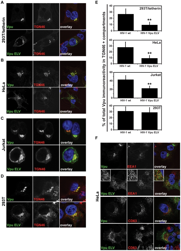
Figure 3. Vpu ELV mutants localize to early endosomal compartments in tetherin-expressing cells.
293T/tetherin (A), HeLa (B), Jurkat (C) or parental 293T (D) were infected with HIV-1 wt or HIV-1 Vpu ELV at an MOI of 1. 48 h later the cells were fixed and stained for Vpu (green) and the TGN marker TGN46 (red) and examined by confocal microscopy. Panels are of representative examples. (E) The percentage of the total Vpu immunoreactivity localized to TGN46+ compartments was calculated for cells (n = 20) from A–D using the Leica Confocal Software. Results were analyzed by unpaired 2-tailed t-test - ** P = 10−8 or lower. (F) HeLa cells as in B were stained for Vpu (green) and the early endosomal marker EEA1 or late endosomal marker CD63 (red).

