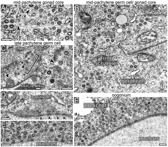
Figure 4. Cer1 capsids localize on microtubules.
(A–F) Electron microscopy of gonads from 15°C adults; regions of the gonad are as indicated at the top of the panels. Capsids localize predominantly to the core in the mid-pachytene region, but concentrate near nuclei in late-pachytene germ cells (C–E) and in oogonia (F). Note that many capsids localize with microtubules (arrowheads) both in the core (panel A) and in germ cells (panel C). P granules are visible in panels C (arrow) and in panel D; arrowheads in panel D indicate examples of nuclear pores. (E) Cluster of capsids near the nucleus of an early oogonium. (F) A “crescent” of capsids by an oogonium nucleus; there are at least 64 capsids and numerous microtubules visible at higher magnifications of this single thin section (data not shown). Scale bars: A–F (0.2 µm).

