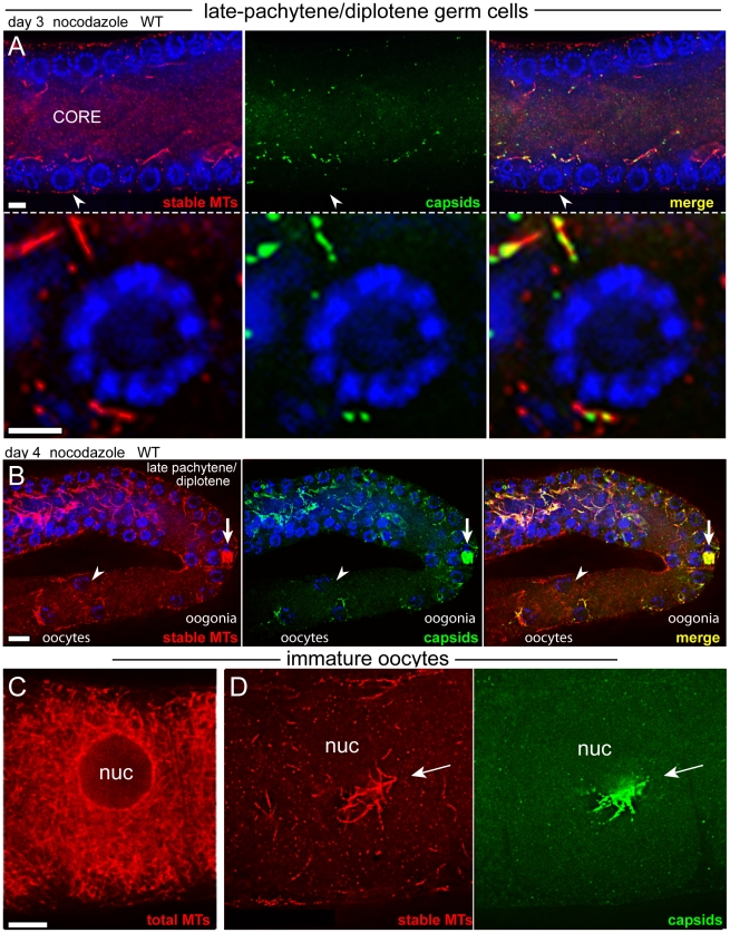
Figure 8. Capsid localization near late-pachytene nuclei requires inhibitor-sensitive microtubules.
(A) Late-pachytene/diplotene region of a nocodazole-treated, day 3 wild-type gonad showing the near absence of stable microtubules and capsids; the arrowhead indicates a germ cell shown a higher magnification in the lower panels. Compare these images with the abundant microtubules and capsids in the same region of untreated gonads (Figure 6A, boxed region, and Figure 5G, respectively). (B) Low magnification image of a nocodazole-treated, day 4 wild-type gonad, oriented as in Figure 6D. Note the rare example of a large aggregate of stable microtubules and capsids in an early oogonium. Because oogonia in this region begin to receive cytoplasmic flow from the gonad core, it is possible that this focus broke off from the mid-pachytene region, rather than surviving the normal developmental progression through late pachytene. Note also that many of the oogonia and early oocytes lack crescents of capsids (arrowhead; compare with Figure 3A), and the few crescents of capsids co-localize with stable microtubules. (C) Untreated, wild-type oocyte showing enrichment of microtubules near the nuclear envelope. (D) Nocodazole-treated, wild-type oocyte showing a focus of stable microtubules and a crescent of capsids. Oocytes shown in both panels occupied the -5 position in the ovulation sequence. Scale bars: A (5 µm), B (10 µm), C (8 µm).

