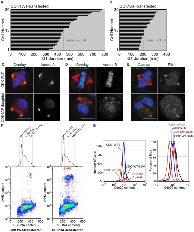
Figure 1. M-phase kinases are present and properly localized on mitotic spindles formed in G1 daughters with impaired APC-Cdh1 activity.
(A) Histogram of G1-phase duration in daughters of CDK1WT-expressing HeLa cells. (B) Histogram of G1-phase duration in daughters of CDK1AF-expressing HeLa cells. (C) CDK1WT M-phase cell and M-phase-like G1 cell (CDK1AF daughter) probed for Aurora A, (D) Aurora B, and (E) Plk1; mCherry-α-tubulin (red) and kinases (green). (F) Pseudo-color scatter plots of DNA content vs. phospho-histone H3 (pHH3) content in 50000 CDK1WT-transfected (left) or CDK1AF-transfected (right) HeLa cells, with DNA content histogram. (G) Histogram of Cdc20 contents in cell cycle phases of CDK1WT-expressing cells and the low DNA-content/high pHH3-content CDK1AF-expressing cells (left); same histogram shown as percent of max (right).

