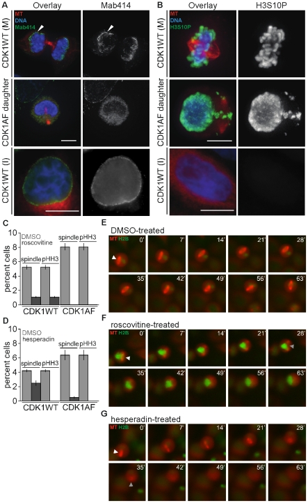
Figure 3. Premature spindle formation in G1 cells requires CDK1 and AurB activities.
(A) Representative mitotic (M) and interphase (I) CDK1WT-expressing cells and a G1 cell produced by division of a CDK1AF-expresser (CDK1AF daughter), presenting microtubules (MT; red), DNA (blue), and nuclear pore complexes (Mab414; green). (B) Different cells from the population in (A), probed for pHH3 content (green). (C) Histogram of percent CDK1WT mitotic or CDK1AF M-phase-like G1 cells with spindles and/or pHH3 content after DMSO or roscovitine treatment. (D) Histogram of percent CDK1WT mitotic or CDK1AF M-phase-like G1 cells with spindles and/or pHH3 content after DMSO or hesperadin treatment. Unsynchronized HeLa cells stably expressing histone H2B-GFP (green) and mCherry-α-tubulin (red) were transfected with CFP-CDK1AF, then 24 h later were treated with DMSO, roscovitine, or hesperadin, and imaged live for 75 min (E–G). (E) M-phase-like G1 cell treated with DMSO. (F) M-phase-like G1 cell treated with roscovitine. (G) M-phase-like G1 cell treated with hesperadin. Scale bars in (A–C) = 10 µM. White arrowheads in (E–G) indicate prematurely formed spindle; gray arrowheads in (F–G) indicate last image of apparent spindle structure.

