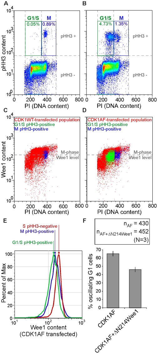
Figure 6. Premature M-phase initiation can be reduced in G1 cells with impaired APC-Cdh1 activation by enforced non-degradable Wee1 expression.
(A) Fifty-thousand CDK1WT-transfected cells and (B) 50000 CDK1AF-transfected cells on a pseudo-color scatter plot of PI (abscissa) and pHH3 content (ordinate). High DNA and high pHH3 content (M phase) gates (blue “M”) and low DNA and high pHH3 (abnormal G1/S cells) gates (green “G1/S”) are shown with their respective frequencies. (C) Cells gated in (A) are overlaid on a pseudo-color scatter plot of PI (abscissa) and Wee1 content (ordinate) of CDK1WT-transfected cells. (D) Cells gated in (B) are overlaid on a pseudo-color scatter plot of PI (abscissa) and Wee1 content (ordinate) of CDK1AF-transfected cells. The M-phase Wee1 level (gray dashed line) is defined in both (C) and (D) by the horizontal axis centered on the Wee1-stained M-phase population that was gated in (A). (E) Histogram of relative Wee1 contents in CDK1AF-transfected cells in normal S phase (pHH3-negative; red), in normal M phase (pHH3-positive; blue), and in precocious M phase (G1-S DNA content, pHH3-positive; green) (F) Histogram of percent oscillating daughters generated in HeLa cells co-transfected with cyclin B1-YFP and CDK1AF alone, or together along with ΔN214Wee1.

