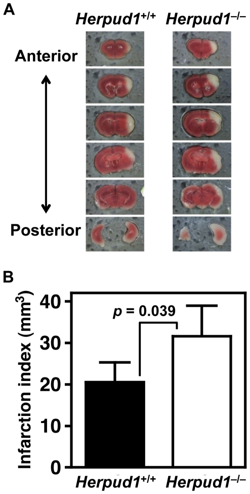
Figure 7. Cerebral infarction models.
Neocortical infarction was induced using the temporary three-vessel occlusion method. Representative images of cerebral infarctions from Herpud1 +/+ and Herpud1 −/− mice stained with 2,3,5-triphenyl-tetrazolium chloride 24 h after ischemia are shown (A). White areas indicate infarct regions. Infarction volumes (n = 6) are expressed as the means with error bars of standard deviation (B).

