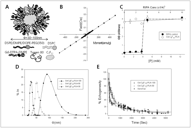
Figure 1. Chemical characterization of Gd-C5F12-PLNs.
(A) Design of Gd-C5F12-PLN. Gd-C5F12-PLN was Φ = 30, 50, and 100 nm. (B) Paramagnetism of the Gd-C5F12-PLN from SQUID test. (C) Cytotoxicity of Gd-C5F12-PLNs in MS-1 cells. The cytotoxicity of Gd-C5F12-PLN was investigated by MTT assay. Serially diluted cell lysis buffer (RIPA) was used as a positive control. (D) Size-distribution of Gd-C5F12-PLNs from DLS analysis. (E) In vitro stability of Gd-C5F12-PLN-50, Gd-C5F12-PLN −100 and SonoVue was calculated from its echogenesity using high-frequency ultrasound.

