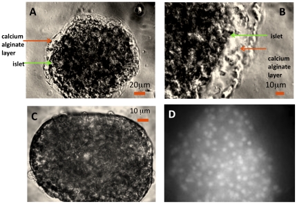
Figure 3. Images of encapsulated islets illustrating the polymerized alginate layer and dye fluorescence.
Islets were placed in the perifusion chamber and brightfield images were taken with a digital camera as described in the Methods section. Images of single islets (A. diameter = 140 µm; B. diameter = 95 µm) coated with transparent calcium alginate. C. Image of unencapsulated islet. D. Fluorescence image of an encapsulated/dyed islet.

